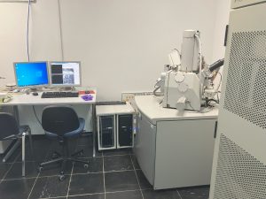
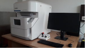
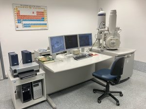
| Field-emission scanning electron microscope FE-SEM JSM-7500F (Jeol) | A high-resolution scanning electron microscope with a cold-field emission source of electrons (cold FEG), which enables observation of the surfaces of sensitive biological samples with nanometer resolution.
|
| Mikro-CT Neoscan N80 | The N80 X-ray micro-tomograph enables X-ray analysis and spatial (3D) reconstruction of native animal and plant specimens. It operates in the range of 40 to 100 keV and achieves a resolution of in a micrometer range. The system is upgraded with a cooling and heating sample stage, allowing observations of frozen objects and analysis of the temperature effect on samples in situ.
|
| AxioImager Z.1 microscope with Apotome system (Zeiss) | The light microscope enables observations by various illumination techniques (bright and dark field microscopy, differential interference contrast (DIC), phase contrast) and epifluorescence. The microscop is upgraded by the Apotome system, which enables 3D and time-lapse analysis of samples.
|
| Motorized fluorescent stereomicroscope SMZ25 (Nikon) | The stereomicroscope enables observation of objects in size range between a few tenths of a millimeter to a few centimeters, isolated animal and plant organs and tissues, bacterial and fungal cultures, and larger tissue slices by various regimes of illumination. These include brightfield illumination by transmitted light, oblique contrast, the dark field illumiation, classic surface illumination and epifluorescence. In addition, stereomicrscope enables image capture without parallax, with motorized movement in the z-axis, as well as image composition with extended depth of field and surface reconstruction in 3D.
|
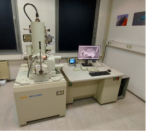
| Field emission scanning electron microscope JSM-7500F (JEOL)
|

1. Sample preparation
- Cryostat (Leica CM3050)
- Vibrato (Precissionary)
- Two-chamber thermo-block for freezing in propane or isopentane cooled with liquid nitrogen
- Lyophilizer (alpha Christ)
- Lyophilizer (ScanVac, Labogene)
2. Data analysis using synchrotron light based techniques:
- Analysis of XRF spectra: Software PyMCA, GeopixeII,
- Analysis of FTIR spectra: Software Opus
- image analysis with multivariate statistical tools (PCA, NNMA): PyMCA, Orange, R-platform
LA-ICP-MS
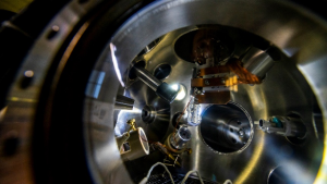
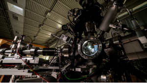
Laser ablation (LA) system (Photon Machines Analyte G2) hyphenated with (a) inductively coupled plasma elemental mass spectrometer (ICP-MS) (Agilent Technologies 7900 ICP-MS) or with (b) inductively coupled plasma time-of-flight elemental mass spectrometer (ICP-TOF-MS) (Nu Ametek, Vitesse).
- list of services for which the equipment is available: (i) 2D/3D elemental (bio)imaging, and (ii) in-situ single-particle (SP) elemental analysis for the needs of nano(bio)metallomics, including sizing and counting, based on the LA-SP-ICP-MS detection mode.
- list of projects/programs related to EuBI/SiMBiON: P1-0034, “Analytics and chemical characterization of materials and processes”.
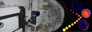
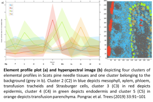
Figure. LA-ICP-MS (left) and illustration of the silver nanoparticle size distribution analysis (right) in root cross-sections of sunflower using LA-single-particle-ICP-MS detection mode.
F5 – Department of Solid-State Physics, Prof. Dr. Igor Serša
100 MHz MRI system for imaging of medium-sized samples and MR microscopy
- 2.35 T horizontal superconducting magnet with a bore of 30 cm
- Tecmag Apollo spectrometer
- gradient system for imaging of medium-sized samples with gradient strength up to 20 mT/m
- RF probe for imaging of medium-sized samples with a diameter of 15 cm
- gradient system for MR microscopy with gradient strength up to 250 mT/m
- RF probes for MR microscopy 10, 15, 20, 25 mm in diameter
- diffusion probe with gradient strength up to 6 T/m
400 MHz MRI system for MR microscopy
- 9.4 T vertical superconducting magnet with 94 mm opening
- Tecmag Redstone spectrometer
- gradient system for MR microscopy with gradient strength up to 800 mT/m
F2 – Department of Low and Medium Energy Physics
X-ray Analytical Microscope (Micro-XRF) Horiba XGT-9000
Micro X-ray fluorescence spectroscopy is an imaging technique based on excitation of the sample with a photon beam of X-ray light of energy 50 keV and diameter 15 m and detection of characteristic X-ray fluorescence rays during raster scanning of the sample. It enables the localization of elements in various solid samples: biological tissues, geological materials, metals, artifacts of cultural heritage. With the XGT-9000 instrument, it is possible to scan samples from a few mm to 15 cm.
Micro-PIXE
Analytical method of excitation of characteristic X-rays during target irradiation with protons PIXE (Proton Induced X-ray Emission) is based on the detection of characteristic X-ray spectral lines emitted by the sample when it is bombarded with protons with energy in the MeV range. Protons are accelerated in a tandem accelerator, and the focused beam is moved in the form of a raster over the selected part of the sample. The beam is focused with quadrupole lenses to a diameter of 700 nanometers (hence the name micro PIXE), this is also the lateral resolution with which elemental or molecular distributions in the sample are measured.
MeV-SIMS
Secondary ion mass spectrometry with primary heavy ions of energies in the MeV range (MeV – SIMS) is a technique used to analyze the molecular composition of solid surfaces and thin films through the use of a focused primary ion beam that knocks secondary ions out of the sample. These are then analyzed through the mass/charge ratio, which is measured with a time-of-flight mass spectrometer. With this, the elemental, isotopic or molecular composition of the surface can be determined to a depth of 1 to 2 nm. The method is particularly useful for imaging of large molecules in biological tissues. Cl-35 ions with an energy of 5.8 MeV are most often used for the primary ion beam, focused down to 700 nm of diameter.
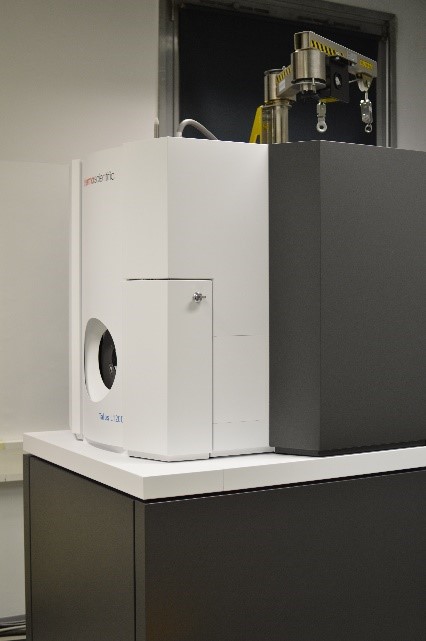
Transmission electron microscope Talos L120C, Ceta camera (Thermo Fisher)
The transmission electron microscope (TEM) (Talos L120C) is specialized for the observation of biological samples, for which its characteristics are optimally adapted, which are high contrast, high resolution at a wide range of magnifications, quick inspection of the image of the preparation on the screen, and capture and processing high resolution images. These characteristics enable the visualization of an extremely wide range of biological samples, i.e. everything from isolated macromolecules, viruses and bacteria to the internal structure of fungal, plant, animal and human cells.
The topics of research and analysis for which the scanning electron microscope Talos L120C is used are extremely diverse, as it is used by a large number of different users for research work in very diverse fields of research, various applications in work for companies, as support for state and government bodies and departments and for pedagogical work.
TEM Talos L120C is majority owned by the National Institute of Biology, while the Department of Biology of the Faculty of Biotechnology of the University of Ljubljana and the Institute of Chemistry are co-owners.
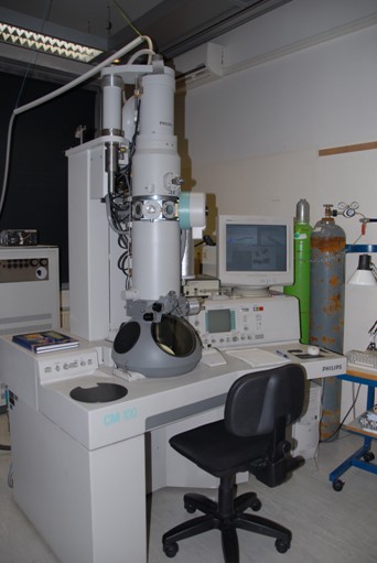
Transmission electron microscope (Philips CM100)
The transmission electron microscope (TEM) (Philips CM100) is equipped with two digital cameras BioScan 792 (Gatan) and Orius SC200 (Gatan) and DigitalMicrograph software for image processing.
The Philips CM 100 transmission electron microscope is successfully used in both basic and applied biological research, as its resolution enables the analysis of cellular ultrastructure. There are two main methods used for research on biological samples. The negative contrast method is suitable for observing particles in suspension, such as viruses, bacteria, macromolecules (proteins, nucleic acids, liposomes). The second method allows observation of plant, animal and human tissues at the nanometer level, i.e. at the level of ultrastructure. It involves tissue fixation, embedding in resins, and cutting ultrathin slices. With the Philips CM 100 TEM, structural analyzes and immunolocalization are performed in various biological samples.
The Philips CM 100 transmission electron microscope is jointly owned by the National Institute of Biology and the Department of Biology of the Biotechnical Faculty of the University of Ljubljana.
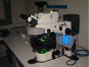
Leica TCS LSI Confocal Stereomicroscope
A confocal stereomicroscope (Leica TCS LSI) enables non-invasive observation of fluorescence in samples. The specialty of this microscope is the wide range of magnifications, as it enables magnifications from 0.7x (16 mm field of view) up to 20x. Due to such a wide range of magnifications, it is possible to observe both larger objects and individual cells. A stereomicroscope also allows for a considerable working distance, which means that larger objects can also be placed under the lens (for example, plants in pots). One of the key advantages of the confocal stereomicroscope over related techniques is the possibility of observing an intact tissue or organ, which shortens the analysis time and significantly increases the relevance of the results.
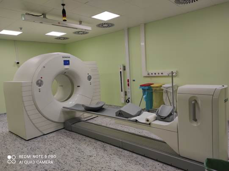
Biograph mCT PET/CT scanner
PET/CT scanner combines the advantages of functional nuclear medicine imaging (PET scanner) and morphological radiology (CT scanner) in a single device. Precise information obtained from the scan leads to a greater possibility of earlier diagnosis and a more accurate treatment strategy, ultimately leading to a better patient outcome.
The design of the Biograph mCT scanner addresses the need for future success. The bore diameter of 78 cm and the tunnel length of 135 cm, as well as the table capacity of 227 kg, allow for the examination of a heavier population and easier patient positioning while offering space for additional therapeutic procedures and helping to increase patient comfort.
The large opening and short tunnel help reduce patient discomfort. The water-cooling component, which reduces the need for noisy fans and over-cooling by air conditioning, creates a calmer environment. In-bed physiological devices and well-placed controls make it easier for technicians to position the patient. The Biograph mCT is designed as a true dual-modality scanner, integrating the best capabilities of PET and CT into one compact system. It was installed in July 2015 with all related equipment.
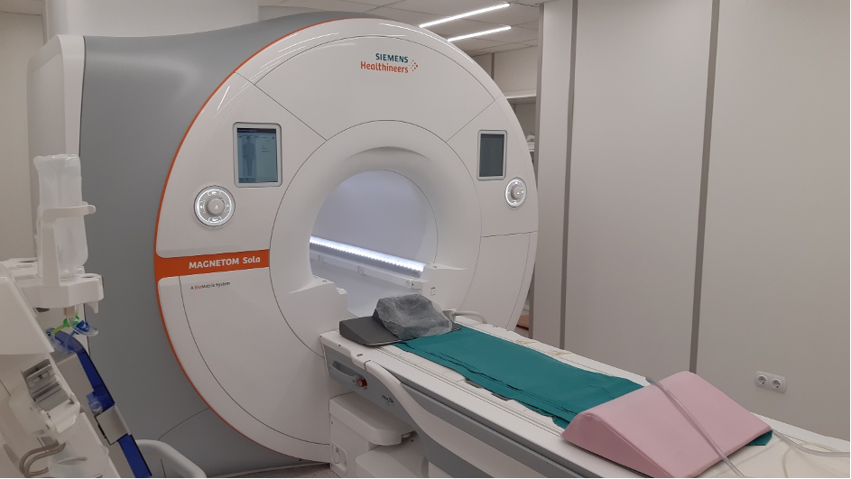
MRI 1,5 T: Siemens Magnetom Sola
The MAGNETOM Sola represents the pinnacle of 1.5T MRI technology, enabling high-quality and individualized examinations. It is the first 1.5T MRI device with biometric technology that automatically adapts to patient biovariability, allowing for higher quality and faster examinations, which is particularly important for oncology patients. The device also enables the optimization of patient appointments and increases work efficiency. It has a wide range of hardware and software options, including a wide range of coils and a pump for contrast application. It was installed in June 2020, when the premises were also renovated.
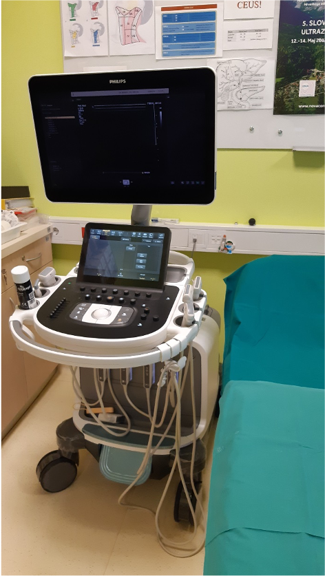
EPIQ 7 Ultrasound Machine
The Philips EPIQ 7 ultrasound machine belongs to the highest class of ultrasound machines. The xMATRIX technology offers all image capture modes (2D, 3D/4D, Live xPlane, Live MPR, MPR, Doppler, Colour Doppler and CPA) in one transducer. MaxVue high-definition display brings extraordinary visualization of anatomy with 1,179,648 additional image pixels compared to a standard 4:3 display format mode, enhances ultrasound viewing and provides 38% more viewing areas to optimize the display of dual, side/side, biplane, and scrolling imaging modes. It has a wide range of probes.
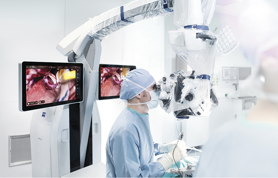
Surgical microscope Zeiss Kinevo 900
KINEVO 900 is an innovative, high-performance robotic visualization system that combines features such as: next-generation motion assistance, on-demand digital 4K visualization capabilities and the QEVO integrated micro-viewing tool. Advanced features include intraoperative fluorescence visualization, an integrated endoscopic examination tool, ocular imaging and a new robotic platform for visualization and three-dimensional (3D) display of images on a 4K monitor, enabling operation in an exoscopic mode.
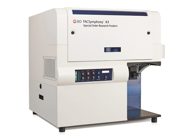
FacSymphony A3 flow cytometer
It is a multi-parameter flow cytometry using an ultra-quiet VPX electronic system, powerful lasers and highly sensitive photomultipliers. It is a powerful tool that allows the scientist to conduct in-depth research. The instrument offers the ability to identify and analyze characteristic phenotypes in heterogeneous populations, analyze 30 parameters in up to five lasers and use new dyes with improved brightness and gradation characteristics.
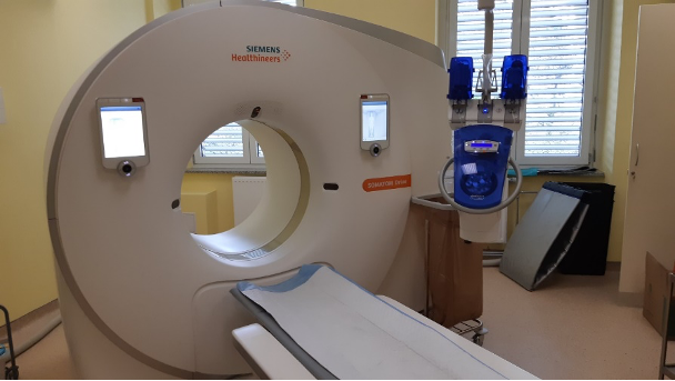
CT Siemens Somatom Drive
It is a second-generation high-performance dual-energy CT machine with two Straton MX Sigma X-ray tubes and two Stellar Infinity detectors. Unique 10 kV tuning levels, the metal filter, Turbo-Flash option, customizable 4D spiral, fast integrated interface with a fast 3D camera, powered by artificial intelligence. It was installed in May 2020, with all the associated equipment, a pump for contrast application, along with the renovation of the premises.
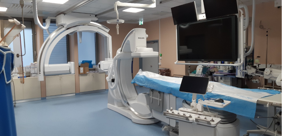
X-ray angiography Philips Azurion
It is a state-of-the-art, latest-generation bi-planar angiography device equipped to perform the most demanding minimally invasive interventional radiology and neuroradiology procedures. It has the latest hardware platform and software. It was installed in June 2020, along with an extensive renovation of the premises and all the related equipment.
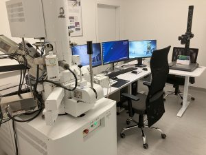
JEOL JSM-IT800SHL
The JEOL JSM-IT800SHL is a multi-purpose FE-SEM offering both high-resolution observation and the capability to conduct various types of analysis, incorporating an in-lens Schottky Plus field emission electron gun (FEG), a Super Hybrid objective Lens (SHL), as well as a new electron optical control mechanism (New Electron Optical Engine: Neo Engine). The electron optical system equipped with an in-lens Schottky Plus field emission gun can deliver a finely focused electron beam even with large irradiation currents, with quickly acquired high accuracy results, including element analysis with EDS, EBSD and Soft X-ray. A high-resolution observation and analysis of a wide variety of specimens from metals, biological specimens to nano materials are possible. The SHL is especially useful for the observation of magnetic materials and analysis such as EBSD measurement. The equipped Soft X-Ray Emission Spectrometer (SXES) is an ultra-high resolution spectrometer consisting of a newly-developed diffraction grating and a high-sensitivity X-ray CCD camera. In the same way as EDS, parallel detection is possible, and 0.3 eV (Fermi-edge, Al-L standard) ultra-high energy resolution analysis can be performed, surpassing the energy resolution of WDS. With the high energy resolution, chemical state analysis mapping can be performed.
FEI Sirion 400 NC
The FEI Sirion 400 NC electron microscope is a high-resolution electron microscope with Field Electron emission, which enables extremely high magnifications (up to a million times, with a high resolution of 1 nm). A feature of this research equipment is its suitability for both engineering materials and biological samples.
FEI Quanta 200 3D
FEI Quanta 200 3D is an environmental Scanning Electron Microscope with a tungsten cathode for an electron source (formation of an electron beam with heat emission). The microscope has the name “environmental” because it allows working at different pressures and at 100% humidity. Conditions for observing different samples are obtained by adjusting the pressure in the chamber. ESEM-mode microscopy is, thus, suitable not only for conductive materials such as metals and metal-coated materials, but also for moist, volatile, and soft materials without prior preparation, which is particularly important for the Food and Chemical industries. Another special feature of this equipment is a focused ion beam FIB SEM (the ion gun on the SEM Quanta microscope), which allows not only SEM observation of the surface, but also surface treatment and analysis of the microstructure below the surface.